IOMP WEBINARS 2022
Radiation Biology Updates: From Low Doses to Ultra-high Dose Rates
Thursday, 15th December 2022 at 12 pm GMT; Duration 1 hour
NEW: CME/CPD credit point shall be awarded for participation in the webinar in full.
To check the corresponding time in your country please check this link:
https://greenwichmeantime.com/time-gadgets/time-zone-converter/
Organizer: Eva Bezak, IOMP
Moderator: M Mahesh, IOMP
Title: Low dose simulation: perspectives in radiation protection
Speaker: Andrea Abril, Profesora Asistente, Departamento de Física, Universidad Nacional de Colombia
Dr Abril is an Assistant Professor and Academic Coordinator for the Master of Medical Physics at Pontificia Universidad Javeriana (Bogotá-Colombia), and since 2022 she is an Assistant Professor (part-time ) at Universidad Nacional de Colombia. She taught subjects such as Advanced radiation physics, Radiobiology, and Numerical Methods for MP which enabled her to integrate her experience in MC simulations with the radiobiological concepts, relevant to imaging optimization. Before that, She held a postdoc position where the main objective was to set up the proof of concept of the development made her my Doctoral thesis (Dr.Sc Universidad Nacional de Colombia 2018 ); a Hybrid device gamma-MRI that integrates dosimetric gel as a gamma radiation detector, the methodologies wrere based on MC simulations.
Abstract:
Doses over 100 cGy have been reported to have evident and consistent biological effects that the patient safety and radiation protection limits are based on it. However, low dose effects are still misunderstood. The NCRP27 compiled relevant studies in which biological responses were reported, even though the effects are unrelated to each other.
The low reproducibility does not permit the determination of the effects observed in the risk models. The main recommendation to boost the knowledge barrier is to design specific experiments at low doses to reproduce the SF curves. DNA mechanistic simulations are a tool to perform in silico experiments under different doses and radiation conditions and to evaluate if they have considerable effects on the SF. These simulations can even distinguish the probability and specific damage reparation mechanism through the damage location to compare with biochemical in vitro assays.
Title: The state of ultra-high dose rate (FLASH) radiotherapy: the rapid developments of a rapid therapy
Speaker: Nolan Esplen, University of Victoria, Canada
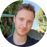
Nolan Esplen is a PhD candidate at the University of Victoria (Victoria, BC, Canada). As a member of the UVic XCITE lab, Nolan’s active research interests pertain to the development of enabling technologies supporting delivery of novel preclinical modalities including spatially-fractionated and ultra-high dose rate (FLASH) radiotherapy. His recent work has found focus on physics and dosimetry within the context of ultra-high dose rate x-ray source development, including that of a megavoltage x-ray platform for FLASH radiobiological research at TRIUMF (Vancouver, BC). Nolan has been awarded a number of honors for his scientific contributions.
Abstract:
Renewed interest in the use of ultra-high dose rates (UHDR) in radiation therapy (RT) has been prompted by a growing body of literature supporting UHDR irradiation, or “FLASH”, as a means to reduce normal-tissue toxicity when compared to irradiation at conventional dose-rates. The potential for improved normal-tissue outcomes (i.e. FLASH effect), paired with isoeffective tumor control and the ability to freeze target motion, has thus made FLASH an attractive candidate for widening the therapeutic window in curative RT. However, while tissue-sparing effects have since been demonstrated across various animal models and radiation modalities (photons, electrons, protons), the radiobiological mechanisms which underlie the FLASH effect, and the beam parameters required to reproducibly elicit it, remain to be elucidated. Continuously updated knowledge has given rise to a number of competing or complementary mechanistic explanations and technologies for more accessible delivery and investigation into this exciting frontier. In this session, a general overview of FLASH radiobiological concepts and delivery techniques will be presented.
PAST WEBINARS 2022
Growing Professional Recognition for Medical Physicists: Raymond Wu and IMPCB
Tuesday, 6th December 2022 at 12 pm GMT; Duration 1 hour
NEW: CME/CPD credit point shall be awarded for participation in the webinar in full.
To check the corresponding time in your country please check this link:
https://greenwichmeantime.com/time-gadgets/time-zone-converter/
Organizer: Tomas Kron, IMPCB Nomination and Election Committee Chair
Moderator: Tomas Kron, IMPCB Nomination and Election Committee Chair
Speakers: Colin Orton, John Damilakis , Adel Mustafa, Ibrahim Duhaini, Art Boyer
(Office bearers and Board members of IMPCB)
Abstract:
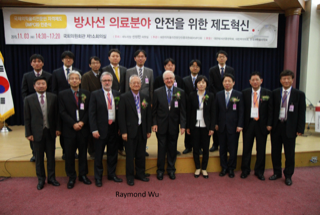
Raymond Wu has been instrumental in setting up the International Medical Physics Certification Board more than 10 years ago. As CEO he has guided the organisation through its rapid growing phase and the many ups and downs typical for an international organisation run by volunteers. IMPCB now has mature processes for accreditation of Medical Physics Certification Boards and certification of medical physicists in countries where no certification board exists. At the end of 2022 Raymond Wu will retire from his position as CEO and the present mini-symposium celebrates his contributions to IMPCB and medical physics.
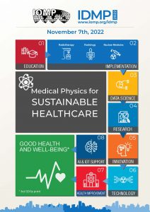
IOMP Webinar on International Day of Medical Physics (IDMP) 2022
Monday, 7th November 2022 at 12 pm GMT; Duration 1 hour
NEW: CME/CPD credit point shall be awarded for participation in the webinar in full.
To check the corresponding time in your country please check this link:
https://greenwichmeantime.com/time-gadgets/time-zone-converter/

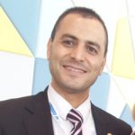
Chairman: John Damilakis, IOMP President
Co-Chairman: Ibrahim Duhaini, IDMP Coordinator
| Invited Speakers | Organization |
| 1. Paddy Gilligan | EFOMP |
| 2. Arun Chougule | AFOMP |
| 3. Patricia Mora | ALFIM |
| 4. Chai Hong Yeong | SEAFOMP |
| 5. Christoph Trauernicht | FAMPO |
| 6. Meshari AlNuaimi | MEFOMP |
| 7. J. Daniel Bourland | AAPM- USA |
| 8. Boyd McCurdy | COMP- Canada |
Joint IOMP-IFMBE Webinar on Clinical Engineering Day 2022: management and maintenance of medical technologies
Wednesday, 19th October 2022 at 12 pm GMT; Duration 1 hour
NEW: CME/CPD credit point shall be awarded for participation in the webinar in full.
To check the corresponding time in your country please check this link:
https://greenwichmeantime.com/time-gadgets/time-zone-converter/
Organizer: Magdalena Stoeva, IOMP
Moderator: Francis Hasford, IOMP
Speakers: Ernesto Iadanza, IFMBE & Magdalena Stoeva, IOMP

Ernesto Iadanza, BME, CE, M. Sc., Ph.D., Senior Assistant Professor in Bioengineering (tenure-track) at the Department of Medical Biotechnologies, University of Siena, nationally qualified as Associate Professor in Bioengineering. He is currently a member of the International Federation for Medical and Biological Engineering (IFMBE) Administrative Council, and Chair of its Council of Societies. He received the IBM Faculty Award in 2013 and the IFMBE/CED Teamwork Award in 2019. Editor in Chief of the Clinical Engineering Handbook 2nd Edition, Academic Press, 2020. Associate Editor of many BME journals. Supervisor in 200+ graduation theses. Author of 190+ publications on international books, scientific journals, volumes, and conference proceedings.
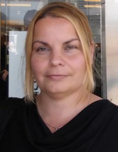
Magdalena Stoeva, PhD, FIOMP, FIUPESM is the present Secretary General of the International Organization for Medical Physics (IOMP) and the International Union for Physical and Engineering Sciences in Medicine (IUPESM) and an Editor-in-Chief of the journal Health and Technology, jointly published by Springer and the IUPESM in cooperation with the WHO.
Dr. Stoeva has expertise in medical physics, engineering and computer systems at academic and clinical level, as well as organizational and international experience.
Prof. Stoeva’s work is directed towards the technological advancements as a driving factor of contemporary healthcare.
IOMP – WHO Joint Webinar on World Patient Safety Day 2022
Wednesday, 14th September 2022 at 12 pm GMT; Duration 1 hour
NEW: CME/CPD credit point shall be awarded for participation in the webinar in full.
To check the corresponding time in your country please check this link:
https://greenwichmeantime.com/time-gadgets/time-zone-converter/
Organizer: John Damilakis, IOMP
Moderator: Eva Bezak, IOMP
Speakers: John Damilakis, IOMP; Emilie van Deventer, WHO; Erik Briers, EPCC

Prof. John Damilakis, MSc, PhD, FIOMP, FIUPESM
John Damilakis is professor and chairman at the Department of Medical Physics, School of Medicine, University of Crete and director of the Department of Medical Physics of the University Hospital of Heraklion, Crete, Greece. He is President of the ‘International Organization for Medical Physics’ (IOMP), Past President of the ‘European Alliance for Medical Radiation Protection Research’ (EURAMED), Past President of the ‘European Federation of Organizations for Medical Physics’ (EFOMP) and Past President of the ‘Hellenic Association of Medical Physics’. Prof. Damilakis is a member of the ICRP Committee 3 and member of the steering committee of the ‘EuroSafe Imaging Campaign’ of the European Society of Radiology. He is coordinator or an active research member of several European and national projects. As a Visiting Professor he has given lectures on dosimetry and radiation protection in Boston University, USA. His publications have been focused on medical radiation protection and dosimetry and, recently, also on Artificial Intelligence in medical imaging. He is editor, author or co-author of several books. Number of publications in PubMed: 243, number of citations: 8286, h-index 48 (Google Scholar). He has received several awards for his work.
Abstract:
Medical Physicists’ Impact on Patient Safety
Medical Physicists are essential healthcare professionals working in varied settings such as hospitals, industry, academia, research institutions, and national radiation protection competent authorities. Regardless of where they work, all medical physicists contribute to safe and quality health care. In radiation safety, a combination of practices and measures are applied to ensure that patients are not exposed unnecessarily, and radiation use is justified. This presentation will summarise the key categories of patient safety issues and provide information about what medical physicists can do to promote patient radiation safety.
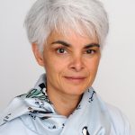
Dr Emilie van Deventer
Dr Emilie van Deventer is the Head of the Radiation and Health Unit at the World Health Organization in Geneva, Switzerland. This programme covers the public health aspects of both ionizing and non-ionizing radiation safety and provides information and guidance to national authorities on radiation protection and health. She is responsible, inter alia, for the WHO International EMF Project, the Global UV project and radon activities. Before joining WHO in 2000, she was a chaired professor of Electrical and Computer Engineering at the University of Toronto, Canada. She holds a PhD from the University of Michigan, USA and an honorary doctorate (doctor honoris causa) from the University of San Marcos, Lima, Peru.
Abstract:
Patient Safety: A WHO perspective
Patient safety is a fundamental principle of health care and is now being recognized as a large and growing global public health challenge. Patient safety is a framework of organized activities that creates cultures, processes, procedures, behaviours, technologies and environments in health care that consistently and sustainably lower risks, reduce the occurrence of avoidable harm, make error less likely and reduce its impact when it does occur. Recognizing the huge burden of patient harm in health care, the 2019 World Health Assembly adopted a resolution on “Global action on patient safety”, which endorsed the establishment of World Patient Safety Day, to be observed every year on 17 September; and recognized “patient safety as a global health priority”. The theme of World Patient Safety Day 2022 is “Medication Safety”, emphasizing the need to adopt a systems approach and promote safe medication practices to prevent medication errors and reduce medication-related harm. In the context of radiation safety, it is important to address quality and safety in the use of radiopharmaceuticals, both in terms of manufacturing and administration/use. (175 words)

Dr ERIK A.M. BRIERS MS PhD
Dr Erik Briers holds a doctorate in Chemistry from the University of Leuven, Belgium. Since his promotion in 1979 he has been involved in laboratory medicine.
He founded in 1982 the diagnostic company Eco-Bio diagnostics and served as its CEO until 1990. In this function he developed a diagnostic test for the detection of Aspergillus antigens with a fast assay allowing the potential curative treatment of patients with disseminated aspergillosis, a deadly disease. This test has been approved by the FDA.
He has been a guest lecturer for the subject “Applied immunology” within the Master study programme Biochemical Engineering at the University of Leuven 2009-2016.
Erik Briers was the Executive Director of the European Cancer Patient Coalition (ECPC) (2012-2013) and ad interim executive director of EPPOSI (The European Platform for Patient Organizations Science and Industry) (2014).
He was active at the European Medicine Agency (EMA) at the Patient and Consumer working party (PCWP) and later was appointed by the EU commission as an alternate patient member of the Committee on Advanced Therapies (CAT) (2016-2019).
He is board member and vice chairman of Europa Uomo.
He is a member of the Guidelines Panel on the treatment of prostate cancer of the European Association of Urology (EAU) since 2013.
He is since 2013 member of the patient advisory board of the European Society of Radiology (ESR-PAG) and has given many presentations at the occasion of the yearly European Congress of Radiology (ECR).
He is vice-president of Us Too Belgium and the chief editor of PROSTAATinfo the magazine of the organization.
He serves as a member of advisory boards of several scientific organizations.
Abstract:
Radiation without harm: Patient’s perspective
Radiation is used in medical applications in imaging, treatment, or as a guiding tool for interventions. The objective is to contribute to the health outcome and the guiding principle is as always “do not harm”.
But radiation by itself can cause harm, depending on the energy and the dose but also on the kind of carrier, photons, electrons etc.
It is a guiding principle that the benefit of a “use” should always outweigh the possible harms or side effects. Thus, when the objective is a screening of apparently healthy individuals for a potentially serious disease such as cancer with a prevalence of some percent the dose should be as low as technically possible. The dose used to treat the discovered cancer on the other hand can be much higher because now we want to harm the cancerous tissue.
The discussion on what is a harmful dose will always depend on the purpose of the application and is to be discussed with the patient, or in case of screening with the healthy individual.
IOMP Webinar: Fractionated radiotherapy and its synergistic relationship with immunotherapy
Tuesday, 21st June 2022 at 12 pm GMT; Duration 1 hour
NEW: CME/CPD credit point shall be awarded for participation in the webinar in full.
To check the corresponding time in your country please check this link:
https://greenwichmeantime.com/time-gadgets/time-zone-converter/
Organizer: Eva Bezak, IOMP
Moderator: Eva Bezak, IOMP
Speaker: Rebecca D’Alonzo, PhD Candidate

Rebecca D’Alonzo, PhD Candidate is a PhD candidate at the School of Physics, Mathematics and Computing, University of Western Australia, Australia. Rebecca is a 3rd year PhD student at the University of Western Australia. She completed her Bachelor of Science at UWA, majoring in Physics and Pathology. She the graduated from Masters of Medical Physics where she won the Master of Physics Medical Physics Prize in 2018. Rebecca is passionate about pre-clinical research, using novel radiotherapy and imaging devices to better understand cancer and how to improve treatment outcomes. Rebecca has also won numerous awards for her research presentations. .
Abstract:
Malignant tumours have decreased oxygenation due to malformed blood vessels. Hypoxia decreases the effectiveness of radiotherapy (RT), and the abnormal vessels prevent both systemic therapies and immune cells from reaching areas of the tumour. This study quantified the alterations of the tumour microenvironment (TME) that can be achieved with varying RT fractionation. The objective was assess the changes in vasculature normalisation and reoxygenation that can be achieved with localised RT. The optimal RT was then combined with various immunotherapy schedules, to find the immunotherapy + RT combination that improves treatment outcomes.
AB1-HA mesothelioma tumour cells were subcutaneously injected into BALB/cJAusbP mice. Mice underwent RT fractionation with a small animal RT device. Starting 10 days post-inoculation, mice received various RT fractionations. Fractions were delivered on consecutive days. On day 15 mice underwent hybrid optical and Doppler ultrasound imaging with a LAZR-X photoacoustic imaging instrument to assess the vasculature and oxygen saturation concentration within the tumour. Imaging continued every second day, until day 29 post-inoculation. The RT fractionation schedule that resulted in the most TME alterations was then combined with various immune checkpoint inhibitor (anti-PD1 and anti-CTLA-4) schedules to find the optimal treatment combination. Mice with cured primary tumour then underwent tumour rechallenge.
Alterations to the TME were observed following different RT fractionation schedules. Imaging showed the most significant increase in vascularisation and oxygen saturation for 2 Gy x 5 fraction. This fractionation schedule was then combined with immunotherapy given at varying timepoints. Immune checkpoint inhibitors given concurrently with RT resulted in the most tumours cured, and lead to tumour rechallenge resistance.
RT fractionation can be used to modulate the TME. This has the potential to be exploited to prime the tumour for susceptibility to other treatments, especially immune checkpoint inhibitors.
Acknowledgements: This study was supported by grant 1163065 from the Cancer Australia Priority-driven Collaborative Cancer Research Scheme.
IOMP Webinar: Non-cancer effects associated with low to moderate doses radiation exposure: what we know so far from epidemiological studies
Monday, 9th May 2022 at 12 pm GMT; Duration 1 hour
NEW: CME/CPD credit point shall be awarded for participation in the webinar in full.
To check the corresponding time in your country please check this link:
https://greenwichmeantime.com/time-gadgets/time-zone-converter/
Organizer: John Damilakis, IOMP
Moderator: John Damilakis, IOMP
Speakers: Marie-Odile Bernier, Sophie Jacob

Marie-Odile Bernier, MD, PhD, works as researcher in the Epidemiology Department of the French Institute for radiation Protection and Nuclear safety (IRSN) since 2005. She is coordinating epidemiological studies on low dose exposure in the field of medical exposure at IRSN. She launched a large cohort specifically designed to study cancer risk in 100 000 children exposed to CT scans in France. She has also more than 25 years of experience as endocrinologist, specialized in the treatment of thyroid cancer.

Sophie Jacob, PhD, is a radiation epidemiologist at IRSN since December 2007. She is specialized in non-cancer effects of ionizing radiation in the context of medical exposure. She coordinated the French O’CLOC study on radiation cataract and lens opacities and was then involved in the OPERRA – EURALOC study (2013-2017), a European multicentric study. In 2013, she initiated research on radiation-induced cardiovascular effects after breast cancer radiation therapy with the French BACCARAT study, further developed in the frame of European MEDIRAD EARLY-HEART study (2017-2022). Dr. Jacob received a PhD in epidemiology (2007) and a master degree in mathematics and biostatistics for biology (2004) from the University of Paris, France.
Abstract:
The use of ionising radiation in medicine is steadily increasing, with clear benefits for population health through improved diagnostic and therapeutic technologies, but it also raises issues in the radiation protection of patients and medical workers.
There is accumulating evidence from epidemiological studies for increased risk of some non-cancer effects following exposure to ionising radiation at low to moderate doses, in particular for circulatory diseases, lens opacities or neurological effects, that may take decades to manifest and present clinically. But there are still uncertainties related to the risks of late-developing non-cancer diseases and effects of radiation exposure.
This webinar will provide an overview of recent epidemiological results and ongoing research in the era of non-cancer diseases related to ionising radiation exposure, with a special emphasis on medical application of radiation.
IOMP Webinar: Computational challenges in patient dose
Tuesday, 10th May 2022 at 12 pm GMT; Duration 1 hour
NEW: CME/CPD credit point shall be awarded for participation in the webinar in full.
To check the corresponding time in your country please check this link:
https://greenwichmeantime.com/time-gadgets/time-zone-converter/
Organizers: Madan Rehani and Pedro Vaz
Moderator: Pedro Vaz, Portugal, Centro de Ciências e Tecnologias Nucleares of IST (University of Lisbon), Portugal.
Speakers: Choonsik Lee, PhD; NCI, NIH, USA & Manuel Bardiès, INSERM (National Institute of Health and Medical Research), France
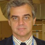
Pedro Vaz, Portugal, Centro de Ciências e Tecnologias Nucleares of IST (University of Lisbon), Portugal.
Pedro Vaz, Ph.D. in Physics, is Coordinator Researcher at Instituto Superior Técnico, University of Lisbon. His areas of research include Radiation Protection and Dosimetry. He was the President of the Center for Nuclear Sciences and Technologies of IST (2017-2020). He is/has been Portuguese Representative in several Committees of the European Union (Group of Experts in Radiological Protection of the EURATOM Treaty and Consultative Committee on Energy Fission of the European Union) and the OECD/NEA (namely the Committee on Radiological Protection and Public Health). He is the institutional representative in European Union research platforms such as MELODI and EURADOS. He served as the National Liaison Officer (NLO) of Portugal for the International Atomic Energy Agency (IAEA). Is member of the Editorial Board of the journal “European Journal of Radiology” and Associated Editor of the journal “Radiation Physics and Chemistry”. Pedro Vaz teaches Radiation Protection and Dosimetry topics in different Portuguese Universities.
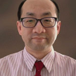
Choonsik Lee, PhD; NCI, NIH, USA
Topic: Current status and challenges in organ dose estimation for patients undergoing diagnostic radiology procedures
Dr. Lee is a senior investigator and Dosimetry Unit head at the United States National Cancer Institute. He has more than 20 years of expertise on computational and experimental radiation dosimetry for patients undergoing medical radiation procedures. His research team develops methods and tools to estimate radiation dose from medical exposures, including treatment and diagnostic tests, in order to generate reliable dosimetry data for use in epidemiological studies of ionizing radiation and cancer risk. Dr. Lee has involved in several task groups in the International Commission on Radiological Protection in the past years and was appointed to the ICRP Committee 2 (Doses from Radiation Exposure).
Abstract
Diagnostic medical radiation sources have substantial contributions to effective dose per capita worldwide. Although radiology procedures provide indisputable benefits to patients, there are still concerns about potential risks from radiation doses, especially in pediatric patients who are more sensitive to radiation than adults. The investigators at the United States National Cancer Institute are developing methods and tools including National Cancer Institute dosimetry system for Computed Tomography (NCICT), nuclear medicine (NCINM), and radiography and fluoroscopy (NCIRF) to estimate individualized organ doses. The tools have been actively used for epidemiological and clinical research. The major outcomes from the research and remaining challenges will be presented at this seminar.
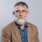
Manuel Bardiès, INSERM (National Institute of Health and Medical Research), France.
Topic: Clinical dosimetry in diagnostics and therapy: recent developments and new perspectives
Manuel Bardiès obtained his Doctorate from Toulouse University, France, in 1991. He developed his research in radiopharmaceutical dosimetry within INSERM (National Institute of Health and Medical Research) since 1992 in Nantes, then in Toulouse, and currently in Montpellier (2021-). He was member of the EANM Dosimetry Committee (2001-2013, chair 2009-2011). He chaired EFOMP Science Committee (2014-2016), and is currently chairing the EFOMP Special Interest Group for radionuclide internal dosimetry. He has been involved in education in various European structures (ESMIT, ESMPE). He’s members of the Board of the medical physics resident programme in France. The group led by Manuel Bardiès is primarily involved in radiopharmaceutical dosimetry, at various scales (cell, tissue, organ). This requires the ability to assess radiopharmaceutical pharmacokinetics, through quantitative imaging. An important part of the research activity involves Monte Carlo modelling of radiation transport. The objective is to improve molecular radiotherapy by allowing patient-specific treatments.
Abstract
Targeted and Selective internal radiotherapy have boosted the development of clinical internal dosimetry. The recent developments include a better appraisal of activity uptake in different compartments of the body, more accurate pharmacokinetics and absorbed dose determination. In addition, there is a growing awareness of the real challenges, both methodological and technological, raised by therapeutic nuclear medicine dosimetry. It is now clear that conventional clinical dosimetry workflows developed for reference dosimetry in diagnostics are not relevant in a context of therapy. More work is needed to better identify the relevant dosimetric indices that will contribute effectively to therapeutic optimisation. On the bright side, the increasing development and dissemination of clinical dosimetry, associated with the advent of commercial treatment planning software participates to both quantitative and qualitative improvements of our professional practice. Now come the time to develop quality assurance in clinical dosimetry.
IOMP Webinar: GEANT4 for medical physics applications: an overview and latest updates & Overview of the Geant4-DNA project
Wednesday, 11th May 2022 at 12 pm GMT; Duration 1 hour
NEW: CME/CPD credit point shall be awarded for participation in the webinar in full.
To check the corresponding time in your country please check this link:
https://greenwichmeantime.com/time-gadgets/time-zone-converter/
Organizer: Eva Bezak, IOMP
Moderator: Eva Bezak, IOMP
Speakers: Associate Professor Guatelli (University of Wollongong) & Prof Sebastien Incerti (University of Bordeaux)
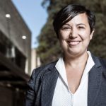
Associate Professor Susanna Guatelli, Centre For Medical and Radiation Physics, University of Wollongong, Australia, is an international leading expert of the Geant4 Monte Carlo code for medical physics applications, working in the field since 2002.
Since 2018, S. Guatelli is member of the Steering Board of the Geant4 International Collaboration in her role of Coordinator of the Geant4 Advanced Examples Working Group. Since 2018, she leads the Geant4 Medical Simulation Benchmarking Group dedicated to the systematic testing of Geant4 for medical applications. S. Guatelli has extensive expertise in teaching Geant4 at various levels and organised many Geant4 short courses addressed to the use of this Monte Carlo code in medical physics.
S. Guatelli has been chair/co-chair of several international workshops and conference sessions dedicated to Monte Carlo codes applied to medical physics. She is Associate Editor of Physica Medica and of Applied Radiation and Isotopes.
In 2021, she was awarded with the prestigious Women in Physics Lecturer award, of the Australian Institute of Physics, for her significant contribution to research at the international level. In 2022, she became a member of the College of Experts of the Australian Research Council (ARC).
Abstract
Topic: GEANT4 for medical physics applications: an overview and latest updates
Geant4, which is a Monte Carlo toolkit describing particle transport and interactions in matter, is widely used in medical physics in critical applications such as verification of radiotherapy treatment planning systems, and the design of equipment for radiotherapy and nuclear medicine. It is also used in medical imaging for dosimetry, to improve detectors and reconstruction algorithms, and for radiation protection assessments.
In this seminar, an overview will be given of the capability of Geant4 for medical physics applications, focusing on the latest updates concerning Geant4 11.0. In addition, she will present the project G4-Med, which has been developed by a large international collaboration, the Geant4 Medical Simulation Benchmarking Group, including both Geant4 developers and users. The goal of the project is to perform a systematic benchmarking of the accuracy of the Geant4 physics models in a set of application scenarios of interest for medical applications.
Information will be provided on how to start to use Geant4 for medical applications. The seminar will finish by describing briefly the Geant4 development processes and Geant4 user support, which are in place to continue to develop this simulation code for and in synergy with the medical physics community.

Professor Sébastien Incerti is director of research at the National Center for Scientific Research (CNRS) and the National Institute of Nuclear and Particle Physics (IN2P3), in France. He is involved in the development of the open source Geant4 Monte Carlo toolkit (http://geant4.org) for the simulation of particle-matter interactions. His research activities focus on the study of the biological effects of ionizing radiation in several application areas, including medical physics and space sciences, in particular for the Geant4-DNA project (http://geant4-dna.org) for which he has been the spokesperson since 2008. Since 2019, he is the scientific director of CNRS-IN2P3 for interdisciplinary science.
Abstract
Topic: Overview of the Geant4-DNA project
In this webinar, we will present an overview of the Geant4-DNA extension (http://geant4-dna.org) of the general-purpose Geant4 Monte Carlo simulation toolkit (http://geant4.org). Initially proposed by the European Space Agency, Geant4-DNA aims at simulating in a mechanistic way the biological effects of ionizing radiation on living organisms at the sub-cellular level. In particular, it allows to simulate the physical, physico-chemical and chemical stages that take place in the biological environment after irradiation, allowing in particular the prediction of early DNA damage from simplified geometries of biological targets. Geant4-DNA is fully included in Geant4 and is freely available to the scientific community. It contains examples of various applications in physics, chemistry and radiobiology, which can be used to learn Geant4-DNA. Its development continues in the framework of an international collaboration.
IOMP Webinar: Virtual imaging trials in breast imaging
Thursday, 12th May 2022 at 12 pm GMT; Duration 1 hour
NEW: CME/CPD credit point shall be awarded for participation in the webinar in full.
To check the corresponding time in your country please check this link:
https://greenwichmeantime.com/time-gadgets/time-zone-converter/
Organizer: John Damilakis, IOMP
Moderator: John Damilakis, IOMP
Speaker: Hilde Bosmans

Hilde Bosmans is professor in medical physics in the University of Leuven (Belgium), she is expert in medical physics in the radiology department of the University Hospital Leuven and steers the physico-medical quality control procedures in 103 mammography units in the Belgian breast cancer screening. She has 261 peer reviewed papers and works currently with 12 PhD students at different themes in medical physics. Research in Virtual Clinical Trials was worked out in the frame of PhD theses by Ann-Katherine Carton, Federica Zanca, Elena Salvagnini, Lesley Cockmartin, Eman Shaheen and Liesbeth Van Coillie. Ongoing projects in VCTs in breast imaging focus on the creation of synthetic data for AI training, the optimization of contrast enhanced mammography and expansion to other modalities such as chest x-ray imaging (with the PhD project of Sunay Rodriguez) and cone beam dental applications.
Abstract
Quality requirements in breast imaging are high, especially in breast cancer screening where the ultimate results are also measured and compared to (European) Guidelines. This has triggered many optimization projects and models for detailed breast dosimetry. The parameter space for optimization is however big, many new techniques are being introduced and clinical trials in breast cancer screening require a huge amount of images and take a lot of time. From the early days on, effort has therefore been put in developing breast models and virtual clinical trial (VCT) frameworks.
We will report on the typical components in VCT studies, on their validation and their limitations. The lecture will conclude with illustrating unique results that could be obtained with virtual imaging trials.
IOMP Webinar: Relative biological effectiveness of protons – time for a change?
Friday, 13th May 2022 at 12 pm GMT; Duration 1 hour
NEW: CME/CPD credit point shall be awarded for participation in the webinar in full.
To check the corresponding time in your country please check this link:
https://greenwichmeantime.com/time-gadgets/time-zone-converter/
Organizer: Madan Rehani, IOMP
Moderator: Eva Bezak, IOMP
Speaker: Iuliana Toma-Dasu
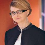
Iuliana Toma-Dasu is Professor in Medical Radiation Physics and the Head of the Medical Radiation Physics division at the Department of Physics, Stockholm University, affiliated to the Department of Oncology and Pathology at Karolinska Institutet in Stockholm, Sweden, and the Editor in Chief of Physica Medica – European Journal of Medical Physics.
Iuliana Toma-Dasu studied Medical Physics at Umeå University, Sweden, where she also became a certified medical physicist and received a Ph.D. degree. In parallel with her involvement in the educational program for the medical physicists run at Stockholm University, her main research interests focus on biologically optimised adaptive radiation therapy, including particle therapy, modelling the tumour microenvironment and the risks from radiotherapy.
Abstract
Current practice in proton radiotherapy planning is based on the assumption that the relative biological effectiveness (RBE) of protons has a constant value of 1.1 as recommended by the ICRU report 78. Nevertheless, increasing evidence is pointing nowadays towards to fact that the RBE of protons is not constant but it varies with the endpoint, the dose per fraction, the actual beam quality described by the linear energy transfer (LET) or other metrics and, of course, the tissue type. This presentation will give a brief overview of the clinical evidence for variable proton RBE and will introduce the frame of the mathematical models for variable RBE based on in vitro cell survival data and the application of these models on proton treatment evaluation and optimisation. A critical discussion on the change from a constant 1.1 proton RBE to a variable value will also be presented.
IOMP-ICRP Webinar: Are radiation risks below 100 mGy for example through recurrent CT procedures of real concern for radiological protection?
Wednesday, 20th April 2022 at 12 pm GMT; Duration 1 hour
NEW: CME/CPD credit point shall be awarded for participation in the webinar in full.
To check the corresponding time in your country please check this link:
https://greenwichmeantime.com/time-gadgets/time-zone-converter/
Organizer: Madan Rehani, IOMP
Moderator: Christoper Clement, ICRP
Speakers: Werner Ruehm, Dominique Laurier, and Richard Wakeford

Christopher Clement is the Scientific Secretary & CEO of the International Commission on Radiological Protection (ICRP), overseeing the daily business of ICRP and representing the organisation in many international fora since 2008. He has presented well over 300 invited lectures in more than 40 countries and overseen the production of more than 70 issues of Annals of the ICRP as Editor-in-Chief. Since 2012 he has been a member of the International Radiation Protection Association (IRPA) Executive Council, and Vice-President of IRPA since 2021. He has more than 30 years of experience in radiological protection, including environmental monitoring and remediation, radiological counterterrorism, and as Director of Radiation Protection at the Canadian Nuclear Safety Commission. In 2019, he received the Ambassador’s Award from the Ambassador of Japan to Canada for his work in recovery after the Fukushima Daiichi accident and the promotion of mutual understanding and friendly relations between Japan and Canada.

Werner Rühm leads the Medical and Environmental Dosimetry Group at the Helmholtz Center Munich, Institute of Radiation Medicine, Germany. In addition, he is professor at the Medical Faculty of the University of Munich. He has been a member of Committee 1 “Radiation Effects” (C1) of the International Commission on Radiological Protection (ICRP) since 2005, serving as C1 Secretary from 2012 to 2016, and as C1 Chair from 2016 to 2021. Currently he is currently chairing ICRP TG91 on Dose and Dose-Rate Effectiveness Factor. From 2014 – 2020 he was Chair of the European Radiation Dosimetry Group (EURADOS), and in 2020 he was elected Chair of the German Radiation Protection Commission (SSK). In 2020 Werner Rühm was elected by the German Federal Parliament and the Federal Council of Germany as a member of the National Civil Society Board, and in 2021 he was elected Chair of ICRP. He is a member of the German delegation to UNSCEAR.

Dominique Laurier has 25 years of experience in the field of radiation epidemiology. He works at the French Institute for Radiation Protection and Nuclear Safety (IRSN) as Deputy Head of the Health Division. He has been a member of the main commission of the International Commission on Radiological Protection (ICRP) since 2017 and chairman of ICRP committee 1 since 2021. He is the French representative to UNSCEAR, and the chair of the Nuclear Energy Agency (NEA) High Level Group on Low Dose Research (HLG-LDR).

Richard Wakeford is Honorary Professor in Epidemiology in the Centre for Occupational and Environmental Health at The University of Manchester, UK. He worked in the nuclear industry for nearly 30 years advising on radiation risks before taking early retirement in 2006, then joining the academic staff of The University of Manchester as Professor before retiring at the end of 2019. Richard has been a member of ICRP Committee 1 since 2009 and has also been a member of a number of expert groups including as a member of the UK delegation to UNSCEAR, the EU Article 31 Group, the UK Committee on Medical Effects of Radiation in the Environment (COMARE), the UK Scientific Advisory Group for Emergencies (SAGE) at the time of the Fukushima Dai-ichi accident, and various technical groups for WHO, IAEA, NEA/OECD and US NCRP. He has been Editor-in-Chief of Journal of Radiological Protection since 1997.
Abstract:
Recent studies suggest that every year worldwide about a million patients might be exposed to doses of the order of 100 mGy of low-LET radiation, due to recurrent application of radioimaging procedures. This webinar provides a synthesis of recent epidemiological evidence on radiation-related cancer risks from low-LET radiation doses of this magnitude. Specifically, reviews of recent results are given with respect to a) the atomic bomb survivors (by W. Rühm), b) low dose-rate exposures during adulthood (by D. Laurier), and c) in utero and childhood exposures. (by R. Wakeford). Taken together, substantial evidence was found that ionizing radiation causes cancer at acute and protracted doses above 100 mGy, and growing evidence for doses below 100 mGy. It is concluded that doses of the order of 100 mGy from recurrent application of medical imaging procedures involving ionizing radiation are of concern, from the viewpoint of radiological protection.
Literature:
W. Rühm, D. Laurier, R. Wakeford, Cancer risk following low doses of ionising radiation – current epidemiological evidence and implications for radiological protection, Mut. Res. – Genetic Toxicol. Environ. Mutagenesis 873 (2022) 503436
M.P. Little, R. Wakeford, S.D. Bouffler, K. Abalo, M. Hauptmann, N. Hamada, G.M. Kendall, Review of the risk of cancer following low and moderate doses of sparsely ionising radiation received in early life in groups with individually estimated doses, Environ. Internat. 159 (2022) 106983
R. Wakeford, J.F. Bithell, A review of the types of childhood cancer associated with a medical X-ray examination of the pregnant mother, Int. J. Radiat. Biol. 97 (2021) 571-92.
IOMP Webinar: Biologically Targeted Radiotherapy: utilising imaging biomarkers to characterise tumour heterogeneity for precision radiation therapy
Tuesday, 22nd March 2022 at 11 am GMT; Duration 1 hour
NEW: CME/CPD credit point shall be awarded for participation in the webinar in full.
To check the corresponding time in your country please check this link:
https://greenwichmeantime.com/time-gadgets/time-zone-converter/
Organizer: Eva Bezak, IOMP
Moderator: Eva Bezak, IOMP
Speaker: Annette Haworth

Prof Haworth is the Director of the Institute of Medical Physics at the University of Sydney and the course coordinator for the medical physics postgraduate program. She has more than 25 years experience as a clinical medical physicist having previously worked at the Peter MacCallum Cancer Centre in Melbourne Australia before moving to Sydney in 2016. Annette’s research interests have focused on novel approaches to brachytherapy and radiotherapy treatments, in particular using quantitative imaging for biological optimization of treatment planning and treatment response.
Abstract:
Intra-tumoral heterogeneity is largely ignored in radiation therapy (RT) treatment planning. Imaging biomarkers derived from quantitative MRI (qMRI) enable voxel-wise mapping of biological characteristics, providing an opportunity to optimise RT dose distributions based on spatially defined Intra-tumoral biology. Mapping changes in qMRI post-treatment offers the opportunity for early identification of those at risk of recurrence. In this presentation I will showcase our work demonstrating how quantitative imaging may be used to produce 3-dimensional maps of tumour heterogeneity to facilitate a precision-based approach to biologically targeted RT treatment planning and treatment response.
IOMP webinar: Image quality monitoring, Medical Physics 3.0, and patient-centered care
Wednesday, 9th February 2022 at 12 pm GMT
NEW: A certificate of attendance will be provided to those who attend the session in full.
To check the corresponding time in your country please check this link:
https://greenwichmeantime.com/time-gadgets/time-zone-converter/
Organizer: Madan Rehani
Moderator: Madan Rehani
Speaker: Ehsan Samei

Ehsan Samei, PhD, DABR, FAAPM, FSPIE, FAIMBE, FIOMP, FACR is a tenured professor, Chief Imaging Physicist, and director of Center for Virtual Imaging Trials at Duke University. Authored over 320 referred papers, he is passionate about bridging the gap between scientific scholarship and clinical practice through virtual clinical trials, clinically-relevant metrologies, and Medical Physics 3.0. He aims towards quantitative patient-centric use of imaging, effectual realization of translational research, and clinical processes that are based on evidence.
Abstract:
While much of medical physics is vested with imaging and therapy technologies, those technologies are effective only to the extend they are used for the care of patients. The physicist is in fact vested with the expertise and the responsibility to ensure that each patient gets the optimum imaging and therapy towards the best clinical outcome. This involves a closer understanding of the clinical nuances of technologies in clinically-relevant quantitative terms, understanding how the advantages of technologies can be effectually realized in its implementation and patient-specific use, and monitoring to ensure the expected outcome is realized. This presentation offers a holistic perspective, anchored to the principles of Medical Physics 3.0, to integrate the rigor of science of medical physics and the relevance of practice towards patient-centered care.
IOMP webinar: Re-igniting the role of physics in medicine
Wednesday, 19th January 2022 at 12 pm GMT
NEW: A certificate of attendance will be provided to those who attend the session in full.
To check the corresponding time in your country please check this link:
https://greenwichmeantime.com/time-gadgets/time-zone-converter/
Organizer: Madan Rehani
Moderator: Madan Rehani
Speaker: Robert Jeraj

Dr. Robert Jeraj is a Professor of Medical Physics, Human Oncology, Radiology and Biomedical Engineering at the University of Wisconsin, Madison and a Professor of Physics at the University of Ljubljana, Slovenia, where he leads international research groups of medical physics. Dr. Jeraj is a founding member of the Topical Group on Medical Physics within American Physics Society (APS), the Working Group on the Future of Medical Physics Research and Academic Training within American Association of Physicists in Medicine (AAPM), the Medical Physics for World Benefit (MPWB) organization, and is a founding Editor in Chief of the Biomedical Physics and Engineering Express (BPEX) journal. He has served as a member of the Medical Imaging Drug Advisory Committee at Food and Drug Administration (FDA), and is on the Board of Commission on Accreditation of Medical Physics Education Programs (CAMPEP). Dr. Jeraj is an author of over 150 published papers, text books and book chapters, and is a frequent invited lecturer and presenter on the use of molecular imaging in therapeutic interventions and general applications of medical physics in radiation and medical oncology.
Abstract:
Medical physics has led to many achievements contributing to modern day medical practice, ranging from high-resolution diagnostics to high-precision treatment deliveries. However, the rapid pace of developments in medicine particularly fueled by personalized therapeutic approaches are posing some unique challenges. At the same time, they are providing new opportunities for involvement of physics in medicine beyond traditional roles. In this lecture, we will (1) review current trends in personalized medicine, (2) summarize some strategic initiatives aimed to address these trends by medical physics societies, (3) highlight the need for additional skills and knowledge required to tackle the challenges and (4) overview some examples of physics involvement in medicine beyond traditional medical physics roles. Specifically, we will present state-of-the-art solutions in the area of quantitative imaging biomarkers, which could be extended and adopted on a global scale.
